Unconventional optical imaging: combining optics-photonics with numeral computing to transcend limits
The field of optical imaging embraces non-invasive imaging techniques using light located on the ultraviolet, visible and infrared bands of the electromagnetic spectrum in order to obtain images of the observed scene or sample. These observed elements are represented through colorimetric greyscale or colour images.
Unconventional imaging enables the access to physical quantity measurements (phase of the sensed light, absorption, refractive index of the sample, polarisation of the illumination wave induced by the observed scene or sample, chemical composition of an object, interaction energy) which are not accessible to conventional measurement systems. Unconventional imaging devices do not directly deliver images, but instead, they rely on specific setups and digital processing of stored data in order to reconstitute an image of the desired physical quantity measurements. This field is also known as computational imaging or quantitative imaging.
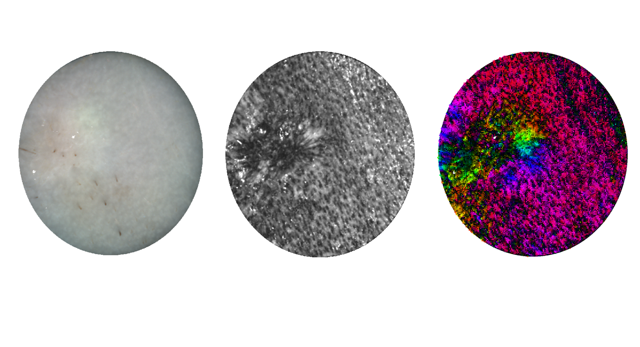
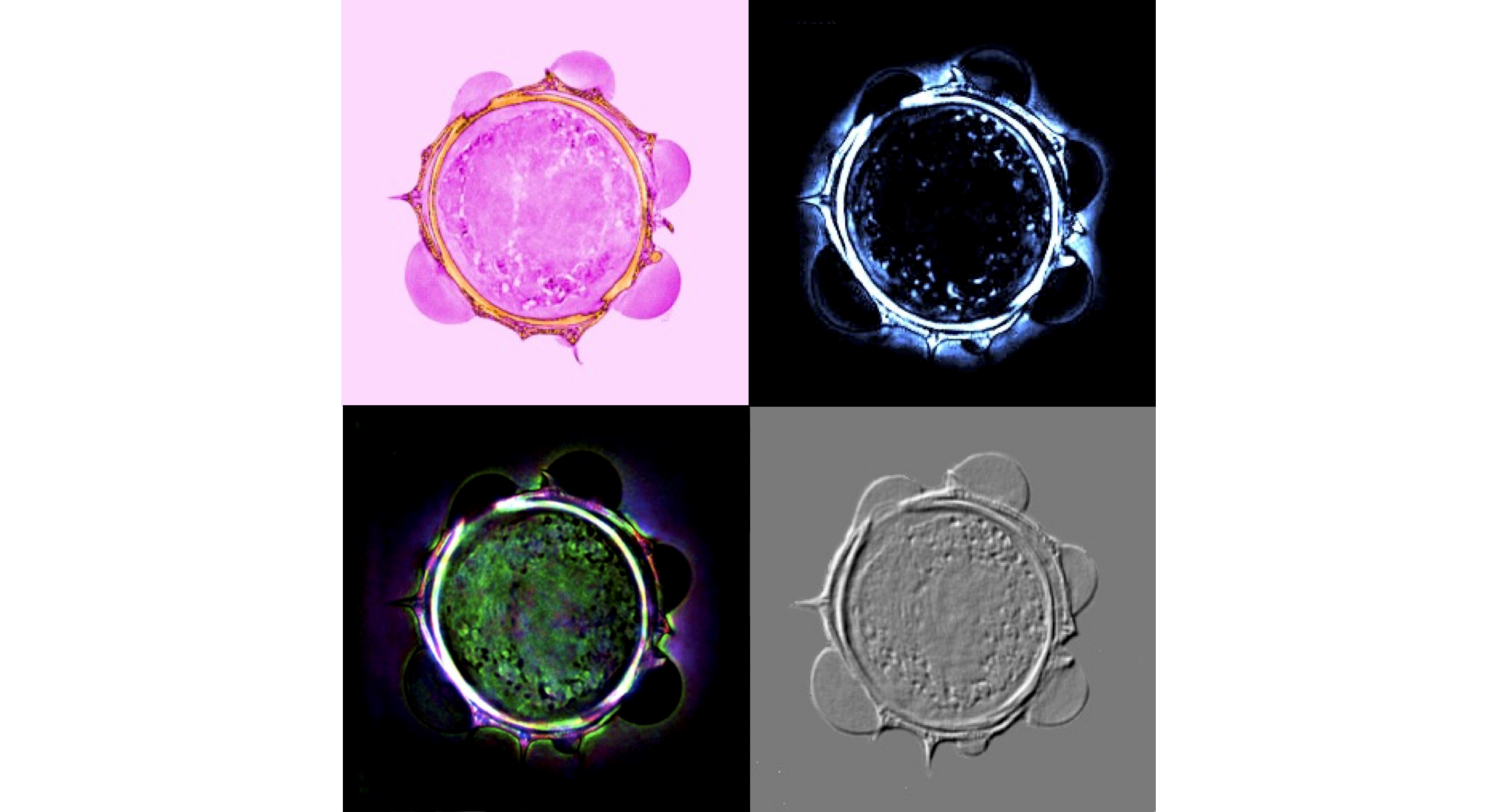
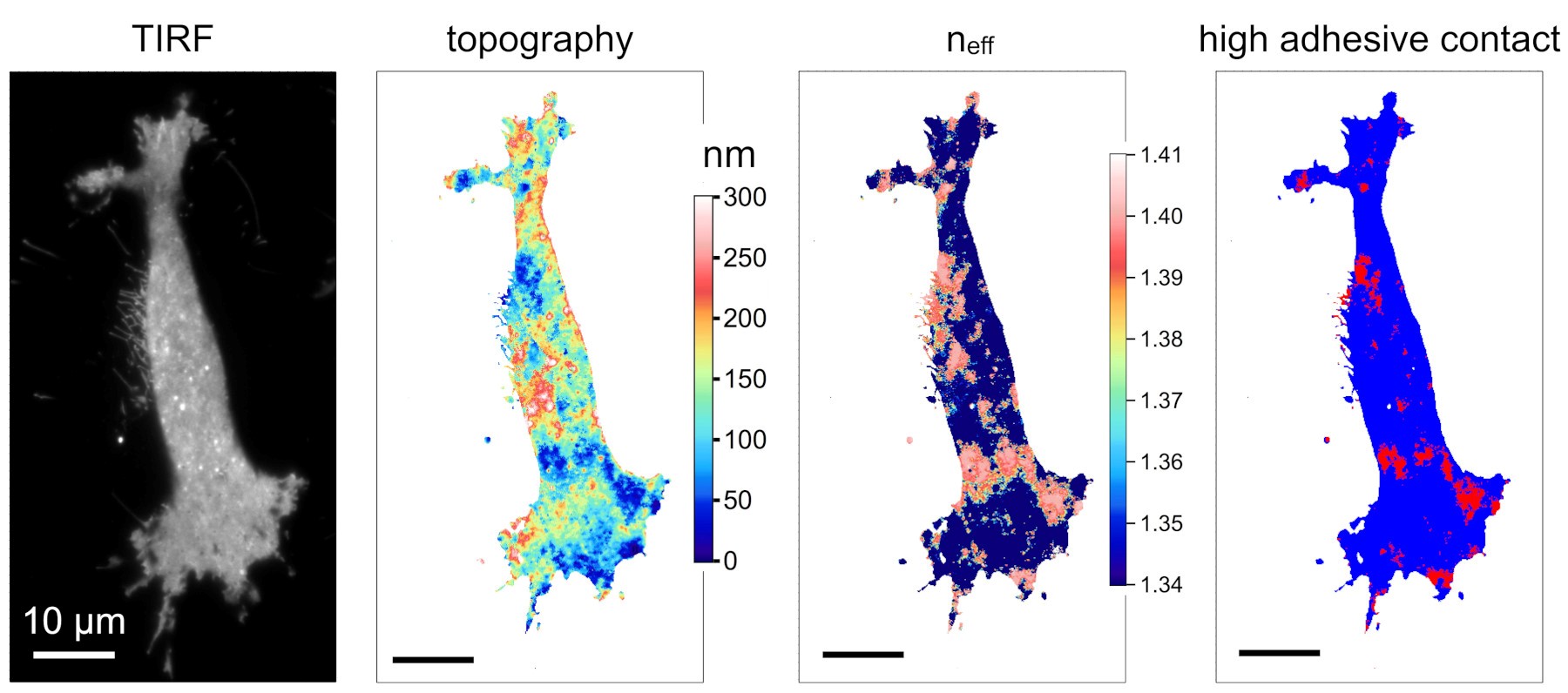
Our innovative developments are very active in the field of optical microscopy, especially in the conduction of research on this area at IRIMAS (holographic microscopy, diffraction tomography imaging), ICube (phase imaging, microsphere-assisted microscopy, local spectroscopy, photonic jet imaging, polarisation imaging) and L2n (Light, nanomaterials and nanotechnologies) (quantitative imaging of cell adhesion).
Our activity also covers the fields of machine vision and quality control, with monocular and Light-Field cameras, with unique innovations in “Light-Field-based deflectometry”, and in robot navigation by camera (visual odometry and trajectory reconstruction via monocular vision). See also the innovation pages.

Other applications

Example of 3D reconstruction of a complex piece by camera-based vision.
Studies in unconventional imaging also find a use in industrial applications. Nowadays, the utilisation of new acquisition modality cameras (event camera, light field cameras) is spreading, with applications in quality control for instance. Robotic imaging specifically allows for effective 3D reconstruction of pieces (IRIMAS – Holo3 partnership). Multidimensional imaging, which includes several modalities (light field and polarimetry for instance) paired with artificial intelligence enables to detect and categorise flaws with high precision.
Raman spectroscopy and sensors
Contexte
Nowadays, the capacity to manage and process multiple data sources to analyse, control and assist in decision making, justifies the use of complex sensors which enables in-real-time acquisition of a great quantity of information, for environmental measures or industrial process control for instance.
Optical sensors, and more specifically spectroscopy-based sensors, enable the in situ in-real-time monitoring of solid, liquid or gaseous materials. The recent technological developments in integrated optics make possible the elaboration of new optical sensors based on Raman scattering.

Description
The innovation consists in using a laser beam focused on the investigated area. This contactless measurement, which can be performed through a transparent container, results in a variable analysis volume ranging from μm3 to mm3. This few-second-long measurement provides several pieces of information: present components and their concentration, flaws in composition or structure, induced or provoked mechanical stress.
The device developed by the LMOPS laboratory is appropriate for many measurement conditions: in a laboratory, paired with another material characterisation technique, chemical or shaping processes on a pilot plant or directly on an industrial system, measurement in full light, outdoor measurement. The aim is to offer an integrated sensor, optimised to provide the desired indicators.
Examples of developed sensors:
Tracking sensors for:
- road salt concentration
- batch or continuous polymerisation reaction
- polymer crystal growth during the shaping process
- polymer spinning process
- fermentation reaction
Chemicophysical characterisation torque sensor:
- rheometer
- X-ray characterisation
- DSC
- tensile testing
- Portable device enabling contactless measurement without prior preparation of the to-be-analysed material
- Remote measurement with optical fibres
- Possible analysis through a non-absorbing interface
- Few-second-long measurement
- Adaptability to conditions and measuring environment
- In situ and ex situ monitoring of chemical synthesis reactions and polymerisation (reagents, products, impurity)
- On-line processes monitoring for shaping materials
- Monitoring of industrial processes
- Monitoring of environment pollutants
The creation process of a Raman-based spectral sensor involves a validation phase within the LMOPS laboratory before its deployment as a field sensor. This validation phase includes a demonstration stage of spectroscopic indicators relevant for measuring followed by a conception and optimisation phase of the optical device as well as data processing optimisation. The development time of the sensor varies according to the desired measurement sensitivity and the constraints of the measurement environment.
- LMOPS – Area of research: photonics
- ICube – Instrumentation et Procédés Photoniques (Photonics Instrumentation and Processes)
Ultra-fast imaging
Numerous physical phenomena occur at infinitesimal time scales challenging our ability to observe them. To capture these fleeting moments, ultrafast cameras prove to be essential. Relying on an advanced expertise in fast electronics and in designing CMOS circuits, the ICube laboratory has developed two families of revolutionary cameras to rise to this ambitious challenge.
The first one, an integrated camera equipped with a CMOS detector makes it possible to image phenomena at the nanosecond scale, revealing details which had been up to that point inaccessible. The second one, a streak camera, pushes the limits even further in allowing for the phenomena to be captured at the picosecond scale. The camera sensitivity goes as far as detecting single photons.
These cameras find many applications in health, in nanochemistry and in monitoring luminous phenomena.
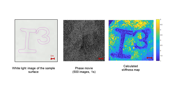
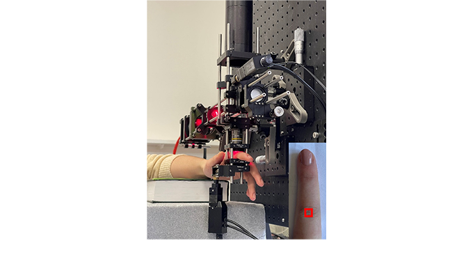
Full-field OCT Elastography
Since the introduction of quantitative elastography in MRI, then in ultrasound scanning, numerous studies have demonstrated the clinical relevance of this contrast for medical imaging, in particular for ultrasound imaging where quantitative elastography is now commonly used for breast cancer screening.
In the Photonics Instrumentation and Processes (IPP) team of the ICube laboratory, we are developing a unique approach stemming from our latest work projects in passive elastography using noise correlation which is currently one of the most promising approaches for in vivo and contactless optical elastography inside biological tissue.
More specifically, the device being developed is a full-field OCT imaging system capable of measuring quantitative mechanical properties with its micrometric 3D resolution while simultaneously providing high-resolution morphological 3D images. This method applies to frequencies ranging from 100 Hz to 10 kHz.
Polarisation imaging
Polarimetric imaging uses the polarisation properties of light to explore physical and biological structures which are invisible when using traditional methods. Its applications include identification of gene signatures specific to cancerous tissue, in-real-time monitoring of their evolution, and measurement of air quality in urban areas. Recently, pairing polarimetric imaging with artificial intelligence has enabled to analyse and make a link between optical variations and biological processes or urban climatic measurement. Furthermore, the advances in quantum polarimetry using entangled photons offer an important opportunity for the conception of photonic components with increased sensitivity and robustness, even in low light. These innovations find promising applications in medicine and teledetection where precision is crucial.