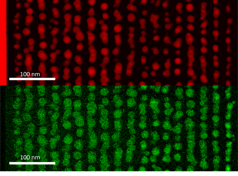
Probing molecular and cellular interactions with ultrafast spectroscopy
Nowadays, understanding the world of living organisms, in particular molecular and cellular interactions, involves the analysis of underlying photochemical mechanisms. We develop femtosecond spectroscopy to study the photoreactivity of (bio)organic or metal-organic molecules in a condensed phase. Photochemical reactions, which enable the conversion of photoelectric or photomechanical energy at the molecular scale, are typical processes of interest. Not only are these photochemical reactions at work in biological systems (photosynthesis, vision), but they also have applications in materials science (photovoltaic devices, molecular motors, photopharmacology). Among interesting photochemical reactions, charge-transfer, photoinduced energy transfer or ultrafast photoisomerisation reactions can be mentioned.
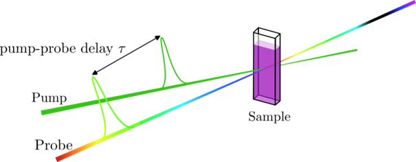
Principle of a pump-probe setup for ultrafast spectroscopy
Quantitative imaging for biomedical applications
To diagnose a pathology or guide a surgical procedure, a simple image is no longer sufficient. The surgeon or histopathologist needs precise quantitative information. Our research work ranges from understanding the interaction between light and biological tissue to its clinical translation from the scale of a cell up to that of an organ. Among the methods that are being developed, the following ones can be mentioned:
- Tilted light-sheet microscopy for 3D imaging;
- OCT (Optical Coherent Tomography) full-field and digital holography for elastography and cancer detection;
- Raman imaging;
- Polarisation imaging;
- Multispectral and hyperspectral imaging for surgical procedures guidance.
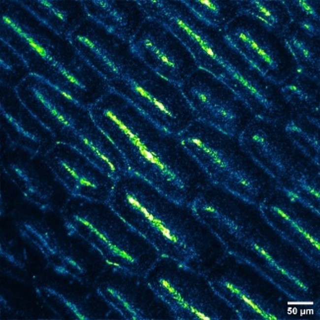
Spider plant leaf through full-field optical coherent tomography (FF-OCT)
Multispectral data acquisition and processing to help diagnose skin cancer
It is now acknowledged that the diagnosis of cancerous lesions at an early stage of development is a key element to increase a patient’s life expectancy. We are working on the implementation of new optical methods to facilitate intraoperative diagnoses, in relation to therapeutic treatment. This process is called theranostics. Ongoing research projects range from the identification of strategic molecular targets to smart data processing algorithms and the development of measuring instruments. Our research activity covers several disciplines related to photonics, multiscale biology, and engineering (instrumentation, signal and image processing, systems). The clinical evaluation of medical devices and innovative software programmes are part of our work.
Publications de référence
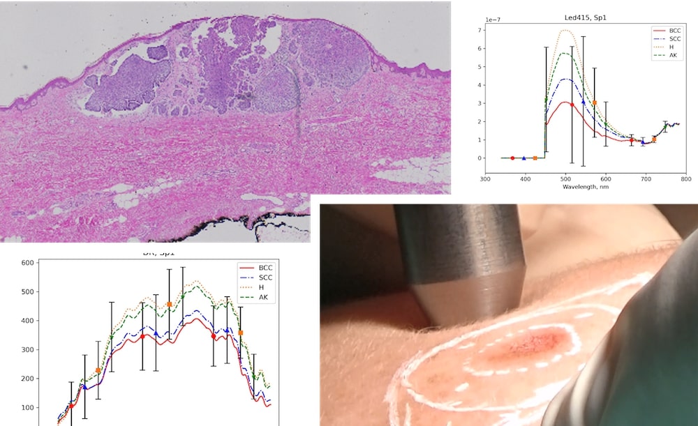
SpectroLive project developed at the CRAN (CNRS-University of Lorraine) in partnership with the CHR (Regional hospital centre) Metz-Thionville, with financial support from the FEDER and the Grand Est Region: photography of a histological cut of a cutaneous carcinoma, example of autofluorescence and diffuse reflection spectrums, photography of the optical probe from the optical spectroscopy device in contact with the skin of a patient included in the clinical study.
Surface plasmon resonance biosensor
Sensors based on fluorescence measurements are nowadays omnipresent in the world of biomedicine, whether utilised for understanding biological mechanisms or for carrying out medical or environmental diagnoses. One of the challenges lies in fluorescence enhancement and the miniaturisation of systems. Our work projects in plasmonics managed to show how these surface or localised light waves enable to concentrate light on the molecules of interest. Our in-depth study projects led to the realisation of highly-integrated prototypes, now commercialised by a spin-off.
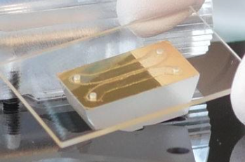
Plasmonic biosensor developed with the spin-off PhaseLab Instrument.
